Mansour H, Azrak R, Cook JJ, Hornburg KJ, Qi Y, Tian Y, Williams RW, Yeh FC, White LE, Johnson GA. The Duke Mouse Brain Atlas: MRI and light sheet microscopy stereotaxic atlas of the mouse brain. Sci Adv. 2025;11(18):eadq8089. Epub 20250430. doi: 10.1126/sciadv.adq8089. PubMed PMID: 40305623; PMCID: PMC12042906.
The Duke Mouse Brain Atlas: MRI and light sheet microscopy stereotaxic atlas of the mouse brain.
MR histology reveals tissue features beneath heterogeneous MRI signal in genetically engineered mouse models of sarcoma.
Blocker SJ, Mowery YM, Everitt JI, Cook J, Cofer GP, Qi Y, Bassil AM, Xu ES, Kirsch DG, Badea CT, Johnson GA. MR histology reveals tissue features beneath heterogeneous MRI signal in genetically engineered mouse models of sarcoma. Front Oncol. 2024;14:1287479. Epub 20240531. doi: 10.3389/fonc.2024.1287479. PubMed PMID: 38884083; PMCID: PMC11176416.
A rapid workflow for neuron counting in combined light sheet microscopy and magnetic resonance histology.
Tian Y, Johnson GA, Williams RW, White LE. A rapid workflow for neuron counting in combined light sheet microscopy and magnetic resonance histology. Front Neurosci. 2023;17:1223226. Epub 20230927. doi: 10.3389/fnins.2023.1223226. PubMed PMID: 37841684; PMCID: PMC10569694.
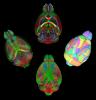
Merged magnetic resonance and light sheet microscopy of the whole mouse brain
Johnson GA, Tian Y, Ashbrook DG, Cofer GP, Cook JJ, Gee JC, Hall A, Hornburg K, Kaczorowski CC, Qi Y, Yeh FC, Wang N, White LE, Williams RW. Merged magnetic resonance and light sheet microscopy of the whole mouse brain. Proc Natl Acad Sci U S A. 2023 Apr 25;120(17):e2218617120. doi: https://www.pnas.org/doi/10.1073/pnas.2218617120 Epub 2023 Apr 17. Erratum in: Proc Natl Acad Sci U S A. 2023 Jun 20;120(25):e2308718120. PMID: 37068254; PMCID: PMC10151475.
Whole-Slide Cytometric Feature Mapping for Distinguishing Tumor Genomic Subtypes in Head and Neck Squamous Cell Carcinoma Whole-Slide Images
Blocker, S. J., Morrison, S., Everitt, J. I., Cook, J., Luo, S., Watts, T. L., & Mowery, Y. M. (2023). Whole-Slide Cytometric Feature Mapping for Distinguishing Tumor Genomic Subtypes in Head and Neck Squamous Cell Carcinoma Whole-Slide Images. Am J Pathol, 193(2), 182–190. https://doi.org/10.1016/j.ajpath.2022.11.004
Prenatal heroin exposure alters brain morphology and connectivity in adolescent mice
Hornburg, K. J., Slosky, L. M., Cofer, G., Cook, J., Qi, Y., Porkka, F., Clark, N. B., Pires, A., Petrella, J. R., White, L. E., Wetsel, W. C., Barak, L., Caron, M. G., Johnson, G. A. (2023). Prenatal heroin exposure alters brain morphology and connectivity in adolescent mice. Nmr Biomed, 36(2), e4842. https://doi.org/10.1002/nbm.4842
Cytostatic hypothermia and its impact on glioblastoma and survival
Enam SF, Kilic CY, Huang J, Kang BJ, Chen R, Tribble CS, Ilich E, Betancur MI, Blocker SJ, Owen SJ, Buckley AF, Lyon JG, Bellamkonda RV. Cytostatic hypothermia and its impact on glioblastoma and survival. Sci Adv. 2022 Nov 25;8(47):eabq4882. https://doi.org/10.1126/sciadv.abq4882
Automated Nuclear Segmentation in Head and Neck Squamous Cell Carcinoma Pathology Reveals Relationships between Cytometric Features and ESTIMATE Stromal and Immune Scores
Blocker SJ, Cook J, Everitt JI, Austin WM, Watts TL, Mowery YM. Automated Nuclear Segmentation in Head and Neck Squamous Cell Carcinoma Pathology Reveals Relationships between Cytometric Features and ESTIMATE Stromal and Immune Scores. Am J Pathol. 2022 Sep;192(9):1305–1320. https://doi.org/10.1016/j.ajpath.2022.06.003
Late-onset cardiovascular dysfunction in adult mice resulting from galactic cosmic ray exposure
Bishawi M, Lee FH, Abraham DM, Glass C, Blocker SJ, Cox DJ, Brown ZD, Rockman HA, Mao L, Slaba TC, Dewhirst MW, Truskey GA, Bowles DE. Late-onset cardiovascular dysfunction in adult mice resulting from galactic cosmic ray exposure. Iscience. 2022 Apr 15;25(4):104086. https://doi.org/10.1016/j.isci.2022.104086
A time-course study of actively stained mouse brains: Diffusion tensor imaging parameters and connectomic stability over 1 year
Xiao, J., Hornburg, K. J., Cofer, G., Cook, J. J., Pratson, F., Qi, Y., & Johnson, G. A. (2022)
A time-course study of actively stained mouse brains: Diffusion tensor imaging parameters and connectomic stability over 1 year. Nmr Biomed, 35(1), e4611. https://doi.org/10.1002/nbm.4611
Restoring morphology of light sheet microscopy data based on magnetic resonance histology.
Tian Y, Cook JJ, Johnson GA. Restoring morphology of light sheet microscopy data based on magnetic resonance histology. Front Neurosci. 2022;16:1011895. Epub 20230104. doi: 10.3389/fnins.2022.1011895. PubMed PMID: 36685227; PMCID: PMC9846533.
Resolution and b value dependent structural connectome in ex vivo mouse brain
Stephanie Crater, Surendra Maharjan, Yi Qi, Qi Zhao, Gary Cofer, James C. Cook, G. Allan Johnson, Nian Wang,
Resolution and b value dependent structural connectome in ex vivo mouse brain, NeuroImage, Volume 255, 2022, 119199, ISSN 1053-8119, https://doi.org/10.1016/j.neuroimage.2022.119199.
A multicontrast MR atlas of the Wistar rat brain
Johnson GA, Laoprasert R, Anderson RJ, Cofer G, Cook J, Pratson F, White LE. A multicontrast MR atlas of the Wistar rat brain. Neuroimage. 2021 Nov 15;242:118470. doi: 10.1016/j.neuroimage.2021.118470. Epub 2021 Aug 12. PMID: 34391877. https://www.sciencedirect.com/science/article/pii/S1053811921007436
Ex Vivo MR Histology and Cytometric Feature Mapping Connect Three-dimensional in Vivo MR Images to Two-dimensional Histopathologic Images of Murine Sarcomas
Blocker SJ, Cook J, Mowery YM, Everitt JI, Qi Y, Hornburg KJ, Cofer GP, Zapata F, Bassil AM, Badea CT, Kirsch DG, Johnson GA. Ex Vivo MR Histology and Cytometric Feature Mapping Connect Three-dimensional in Vivo MR Images to Two-dimensional Histopathologic Images of Murine Sarcomas. Radiol Imaging Cancer. 2021 May;3(3):e200103. doi: 10.1148/rycan.2021200103. PMID: 34018846; PMCID: PMC8183263. https://doi.org/10.1148/rycan.2021200103
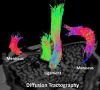
A high-resolution interactive atlas of the human brainstem using magnetic resonance imaging
Conventional atlases of the human brainstem are limited by the inflexible, sparsely-sampled, two-dimensional nature of histology, or the low spatial resolution of conventional magnetic resonance imaging (MRI). Postmortem high-resolution MRI circumvents the challenges associated with both modalities. A single human brainstem specimen extending from the rostral diencephalon through the caudal medulla was prepared for imaging after the brain was removed from a 65-year-old male within 24 h of death. The specimen was formalin-fixed for two weeks, then rehydrated and placed in a custom-made MRI compatible tube and immersed in liquid fluorocarbon. MRI was performed in a 7-Tesla scanner with 120 unique diffusion directions. Acquisition time for anatomic and diffusion images were 14 h and 208 h, respectively. Segmentation was performed manually. Deterministic fiber tractography was done using strategically chosen regions of interest and avoidance, with manual editing using expert knowledge of human neuroanatomy. Anatomic and diffusion images were rendered with isotropic resolutions of 50 μm and 200 μm, respectively. Ninety different structures were segmented and labeled, and 11 different fiber bundles were rendered with tractography. The complete atlas is available online for interactive use [insert link to https://civmvoxport.vm.duke.edu/voxbase/login.php?return_url=%2Fvoxbase%2F]. This atlas presents multiple contrasting datasets and selected tract reconstruction with unprecedented resolution for MR imaging of the human brainstem. There are immediate applications in neuroanatomical education, with the potential to serve future applications for neuroanatomical research and enhanced neurosurgical planning through “safe” zones of entry into the human brainstem. https://doi.org/10.1016/j.neuroimage.2021.118135
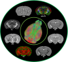
Variability and heritability of mouse brain structure: Microscopic MRI atlases and connectomes for diverse strains
Wang N, Anderson RJ, Ashbrook DG, Gopalakrishnan V, Park Y, Priebe CE, Qi Y, Laoprasert R, Vogelstein JT, Williams RW, Johnson GA. Variability and heritability of mouse brain structure: Microscopic MRI atlases and connectomes for diverse strains. Neuroimage. 2020 Nov 15;222:117274. doi: 10.1016/j.neuroimage.2020.117274. Epub 2020 Aug 18. PMID: 32818613; PMCID: PMC8442986. https://www.sciencedirect.com/science/article/pii/S1053811920307606?via%3Dihub
Cytoarchitecture of the mouse brain by high resolution diffusion magnetic resonance imaging
Characterization complex collagen fiber architecture in knee joint using high-resolution diffusion imaging
Wang, Nian, Anthony J. Mirando, Gary Cofer, Yi Qi, Matthew J. Hilton, and G Allan Johnson. “Characterization complex collagen fiber architecture in knee joint using high-resolution diffusion imaging.” Magn Reson Med, January 21, 2020. https://doi.org/10.1002/mrm.28181.

- Need help? 919-684-7755