DUKE TODAY - DURHAM, N.C. – Magnetic resonance imaging (MRI) is how we visualize soft, watery tissue that is hard to image with X-rays. But while an MRI provides good enough resolution to spot a brain tumor, it needs to be a lot sharper to visualize microscopic details within the brain that reveal its organization.
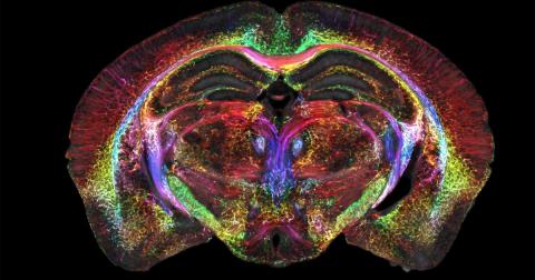
In a decades-long technical tour de force led by Duke’s Center for In Vivo Microscopy with colleagues at the University of Tennessee Health Science Center, University of Pennsylvania, University of Pittsburgh and Indiana University, researchers took up the gauntlet and improved the resolution of MRI leading to the sharpest images ever captured of a mouse brain.
Brain Images Just Got 64 Million Times Sharper | Duke Today
today.duke.edu